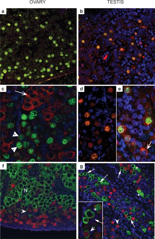
Figure 1:
Distinct subcellular localization of OCT-3/4, DAZL and VASA in human fetal gonads.
In contrast to mouse gonocytes, which express Oct-3/4, Dazl and Mvh proteins, first trimester ovaries (a, 61 day), and testes (b, 64 day) express only OCT-3/4 (green) and DAZL (red) both of which co-localize to germ cell nuclei. In ovaries from the second trimester (c, 14 week) DAZL protein is almost exclusively cytoplasmic and largely localizes to OCT-3/4-negative groups of cells (arrow); a few OCT-3/4-positive cells display low levels of DAZL expression in their cytoplasm (arrowheads). In second trimester testes (d, 16 week; e, 19 week), DAZL is still expressed in the nuclei of some OCT-3/4-positive germ cells but this pattern of expression is variable, with DAZL protein present in the cytoplasm of OCT-3/4-positive and OCT-3/4-negative (arrow, e) cells. (f) Mutually exclusive expression of OCT-3/4 and VASA in the human fetal ovary; cells with intense immuno-positive staining for OCT-3/4 are found at the periphery of the organ (red nuclei), while cytoplasmic VASA protein is most intense in cells located in nests (N) closer to the centre of the ovary. An intermediate population of cells with low intensity nuclear staining for OCT-3/4 and low intensity staining for VASA (arrowheads) was also present. (g) In second trimester human fetal testis, OCT-3/4-positive and VASA-positive germ cells are found within the same seminiferous cords; germ cells with intense nuclear OCT-3/4 expression (red nuclei) are VASA-negative and those with intense cytoplasmic expression of VASA (e.g. arrowed in inset g) were OCT-3/4-negative. Two other populations of male germ cells can be identified; a population with low intensity expression of both OCT-3/4 and VASA (arrowheads) and cells with nuclear VASA expression (asterisks) which were typically found in pairs (adapted from Anderson et al. (2007).

