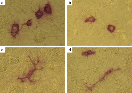
Figure 2:
Morphology of human fetal gonocytes in vitro.
When cultured in vitro on feeder layers of mitotically inactivated STO cells, gonocytes isolated from first trimester testes or ovaries (∼60–65 days gestational age) adopt one of two morphologies, namely round (a, b and d) or migratory (c and d). Both populations can be found in close proximity (d). Germ cells, but not feeders, display strong alkaline phosphatase activity (red).

