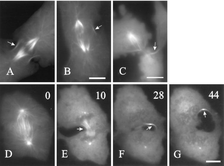
Figure 7.
Correlation between microtubule bundles and furrowing. Cells were treated with ICRF-187 and microinjected with TAMRA-tubulin as in Figure 5. Panels A–C show cortical microtubule bundles in three separate cells. Microtubule bundles were always associated with the ingressing margin, including those misplaced (B, arrow) or formed ectopically (C, arrow). A similar situation was observed when cells were treated with nocodazole during anaphase. Panels D–G show a sequence taken from a cell 0, 10, 28, and 44 min after the addition of nocodazole. Often a small number of microtubule bundles persisted and remained associated with the furrow along a twisted path (arrows). Bars, 20 μm.

