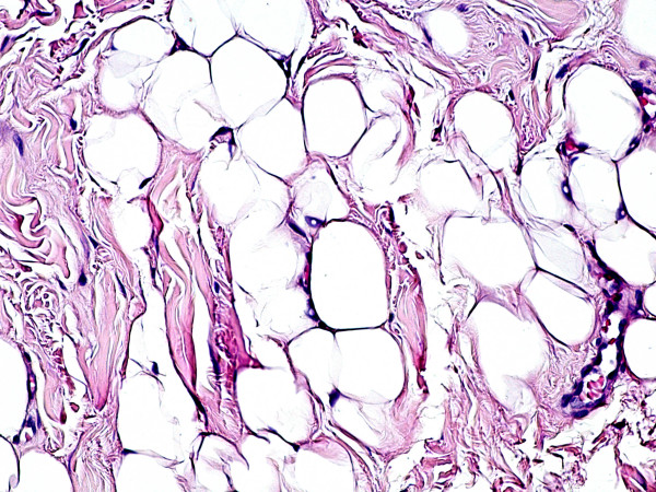
Figure 3.
Histological features of fibrolipoma at high-power magnification. The tumour is composed of mature and univacuolated fat cells, embedded in dense collagen fibres. No morphological or structural alterations of the tissues due to the thermal cut of the diode laser are detectable (haematoxylin-eosin stain, original magnification ×20).

