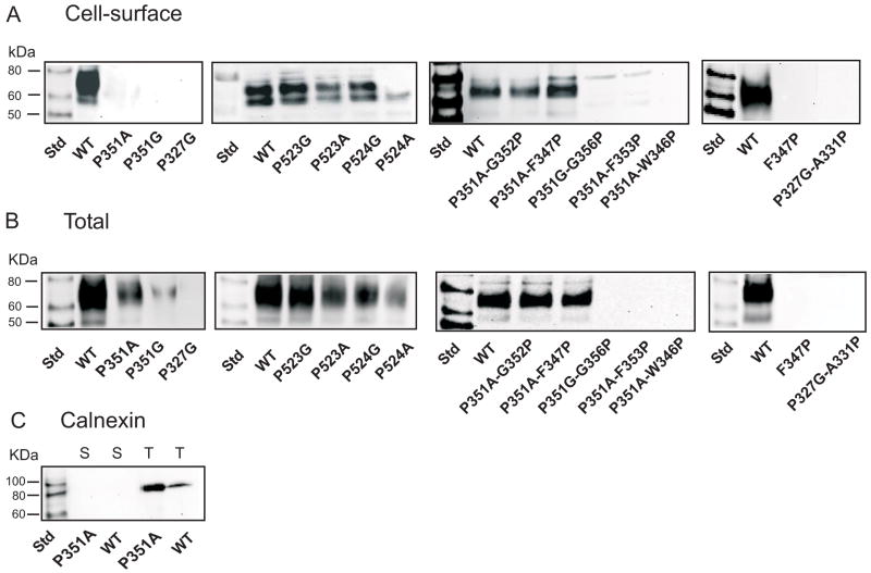
Figure 2.
Western blots of (A) cell surface and (B) total biotinylated protein expression. HRPE cells were transiently transfected with wild-type and mutant rbNaDC1 and then treated with Sulfo-NHS-LC-biotin. For total protein expression, cell lysis buffer was added before addition of Sulfo-NHS-LC-biotin. Western blots were probed with 1:1000 dilution of anti-NaDC1 antibodies, followed by 1:5000 dilution of horseradish peroxidase-linked anti-rabbit Ig. The two bands represent differently glycosylated forms of rbNaDC1 (6;22). (C) Western blots of cell surface (S) and total (T) biotinylated proteins probed with anti-calnexin antibodies (1:2000 dilution), as a marker of endoplasmic reticulum. Chemiluminescent molecular weight standards (Std) are shown in first lane of each blot.

