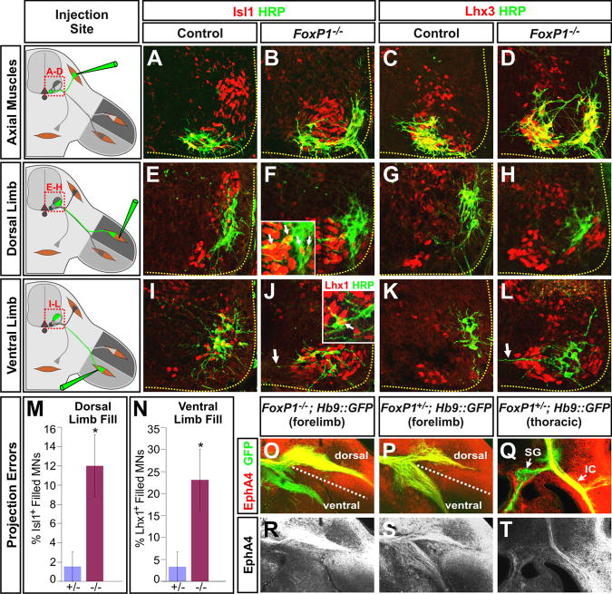
Figure 5. Topographic misprojections of motor axons in Foxp1 mutant embryos.
(A-D). MN projections to axial muscles were traced using HRP injections into axial muscles in e13.5 control and Foxp1 mutant embryos, and subjected to costaining analysis with the indicted antibodies.
(E-H) MN projections to the dorsal limbs were similarly traced using HRP injections. In both control and Foxp1 mutants, most dorsal projecting MNs lacked Isl1 staining and instead expressed Lhx1 (data not shown). However, in the Foxp1 mutants, some of the dorsally projecting neurons aberrantly expressed Isl1 (inset in panel F).
(I-L) Injections of HRP into the ventral limbs labels a dorsolaterally positioned group of Isl1+ cells in the controls, and a ventromedially positioned group of Isl1+ MN in Foxp1 mutants. Some ventrally projecting MNs in the Foxp1 mutants express both Lhx1 and Isl1 (inset in panel J). Arrows in panels J and L indicate the unusual horizontal morphology of dendrites labeled by retrograde labeling from the ventral limbs in the Foxp1 mutants.
(M) Quantification of retrograde labeling of MNs following HRP injections into dorsal and ventral limb muscles. The percentage of HRP labeled MNs that are Isl1+ following injections into the dorsal limb or Lhx1+ following injections into the ventral limbs are shown (p < 0.05 in both cases).
(N, O, Q, R) Distribution of EphA4 receptor in vibratome sections of the rostral brachial plexus from control or Foxp1-null littermates.
(P, S) Equivalent analysis of EphA4 expression in thoracic sections of wild-type embryos. SG, sympathetic ganglia; IC, intercostal nerves.

