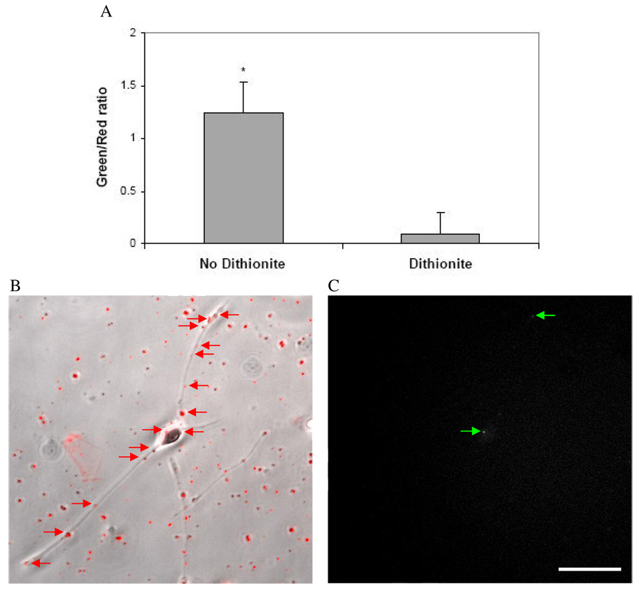
Figure 6. Ratiometric analysis distinguishes between intracellular and extracellular lipoplexes in neuron-like PC-12 cells.
(A) The ratio of NBD to TR fluorescence signals is significantly different between dithionite-treated and untreated particles (*p < 0.001). NBD/TR-double-labeled lipoplexes that associated with the cell are indicated by red arrows in the brightfield/fluorescence overlay image (B), while lipoplexes that were internalized retained NBD fluorescence and are indicated by green arrows (C). (Scale bar = 50 µm)

