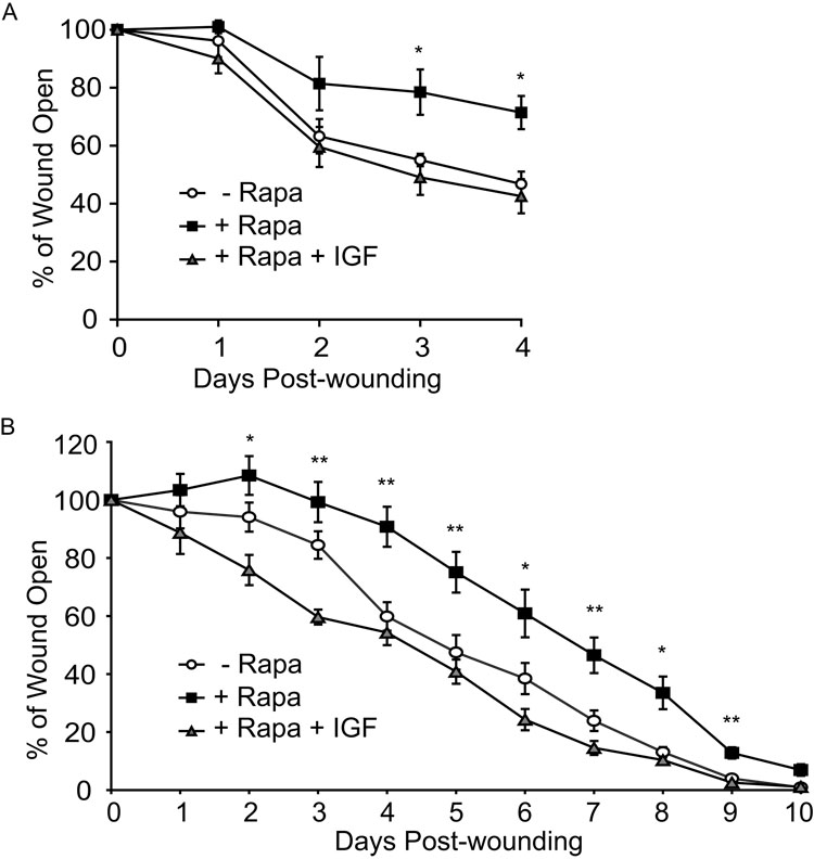
Figure 7. Application of IGF-1 to wounds rescues rapamycin-induced wound healing defect.
(A) IGF-1 supplemented to rapamycin-treated skin in culture rescues wound closure. Skin from wildtype mice was wounded and cultured in the presence or absence of rapamycin. Digital images were acquired to monitor wound closure. 100ng/ml IGF-1 was supplemented in various wells. (B) IGF-1 rescues wound repair in rapamycin-treated mice. Mice were treated with rapamycin or vehicle control, wounded on the dorsal surface, administered IGF-1 or buffer control, and wound closure measured over time. Data represents mean ± SEM. *P < 0.05, **P < 0.005 versus vehicle control (2-tailed, unpaired Student’s t test).

