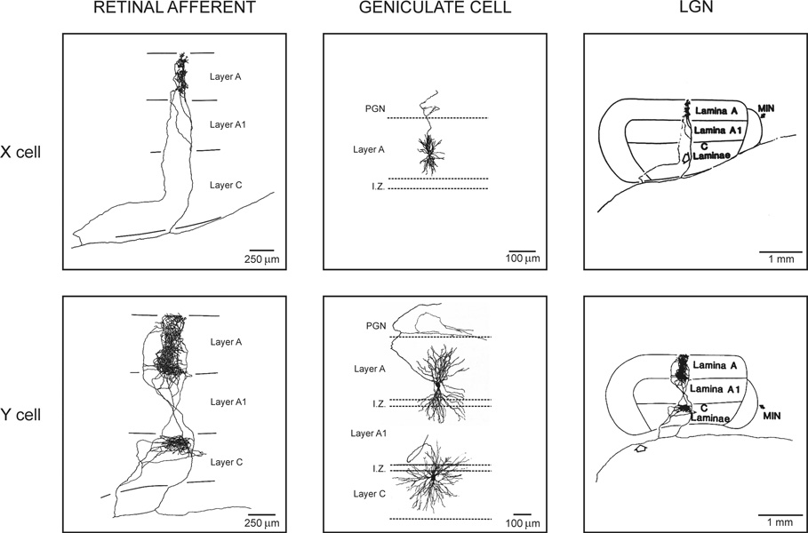
Figure 1. Retinal afferents and geniculate cells.
Left. Axon terminals from X and Y retinal afferents in LGN. X retinal axons project into a single LGN layer and they are very restricted. Y retinal axons can project into two different LGN layers and are wider. Middle. X and Y geniculate cells. X cells have small dendritic trees that are restricted to a single LGN layer. Y cells have larger dendritic trees that frequently cross layers. Right. The same axon terminals from the left of the figure shown at a different scale. Reprinted with permission from (Sur & Sherman, 1982, Copyright 1982 AAAS; Sur, 1988) (left and right) and (Friedlander et al., 1981) (middle).

