
Figure 3.
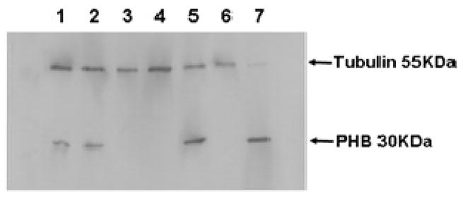
Purity of the mitochondrial preparation. 3 μg of protein from each fraction during purification was subjected to electrophoresis. Proteins were transferred to PVDF membrane and probing with antibodies directed against tubulin as a cytoskeletal marker and prohibitin as a mitochondrial marker. Lanes contained the following fractions: 1. Total homogenate; 2. Post-nuclear supernatant; 3. Nuclear pellet; 4. Post-mitochondrial supernatant; 5. Crude mitochondrial pellet; 6. Microsomal fraction; and 7. Purified mitochondria.