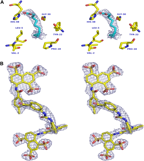
FIGURE 5.
Fo-Fc map shows ligand in ecarpholin S structure. (A) Fo-Fc map in hydrophobic channel of ecarpholin S. Bound lauric-acid molecules were omitted before refinement and map calculation. Map was contoured at a level of 2σ. (B) Fo-Fc map for suramin complex. Inhibitor suramin molecules were omitted before refinement and map calculation. Map was contoured at a level of 2σ. All suramin molecules of asymmetric unit have the equivalent quality of electron density; one representative molecule is shown here. Images were prepared using the program Pymol (59).

