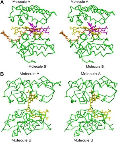
FIGURE 7.
Stereo diagram of comparison of suramin bound to ecarpholin S and Basp-II. (A) Three suramin molecules are sandwiched between monomer A and monomer B of ecarpholin S. (B) In Basp-II/suramin complex (Protein Data Bank code, 1Y4L), only one suramin molecule is located between the two monomers. Image was prepared using the program Pymol (59).

