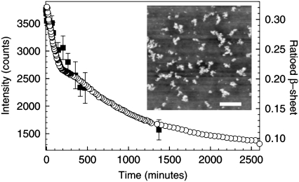
FIGURE 2.
Light scattering and FTIR studies of the disintegration of prefibrillar aggregates in solutions of BPI (20 mg mL−1, pH 1) after a temperature quench from 60°C to 25°C. Data are shown for the decay in the total light scattering intensity (▪, DLS) and the area of the ratioed β-sheet peak (○, FTIR, see text) as a function of time. The inset shows an AFM image of clusters of prefibrillar aggregates taken over a 5 μm × 5 μm area. The scale bar on this image corresponds to a distance of 1 μm.

