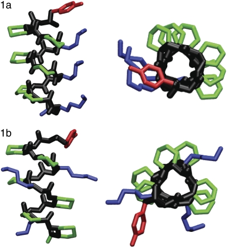
FIGURE 3.
Stick representations of β-peptides 1a and 1b considered in this work. The figures are colored with the backbone in black, the cyclic residues in green, the β3-homolysine residues in blue, the β3-homotyrosine residue in red, and the β3-homophenyalanine residues in red. For clarity, hydrogens have been removed. On the left is a side view and representation on the right is shown looking down the helical axis. These figures were made using VMD (77).

