
Figure 6.
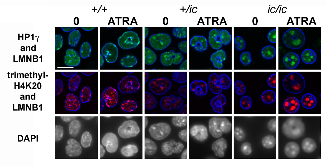
Confocal immunostaining of undifferentiated and granulocytic EPRO cells with anti-HP1γ, anti-me3H4K20 and anti-lamin B1. Genotypes: wildtype, +/+; heterozygous ichthyosis, +/ ic; homozygous ichthyosis, ic/ ic. Cell states: 0, undifferentiated; ATRA, granulocytic forms on day 4. Stains: anti-HP1γ (green); anti-me3H4K20 (red); anti-LMNB1 (lamin B1, blue); DAPI (DNA, uncolored). Fixation: PFA. Scale bar: 10 µm.