
Fig. 4.
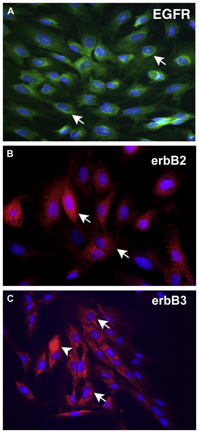
Localization of the ErbB receptors in cultured goblet cells. Localization of the EGFR (green) is shown in (A). Arrows indicate perinuclear staining of cultured goblet cells. Localization of erbB2 (red) is shown in (B). Arrows indicate cytoplasmic staining in cultured goblet cells. Localization of erbB3 (red) is shown in (C). Arrows indicated cytoplasmic staining in cultured goblet cells. Arrowheads indicate cells in which staining is concentrated throughout the cytoplasm. In all micrographs, the nuclei were counterstained with DAPI and are shown in blue. Micrographs are representative of 3 individual animals. Magnification ×400.