
Fig. 8.
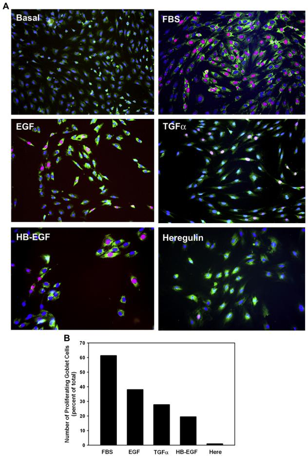
Effect of EGF family of growth factors on goblet cell proliferation. Goblet cells from primary culture were stimulated with either no addition, the positive control FBS (10%), EGF (10−7 M), TGF-α (10−7 M), HB-EGF (10−7 M), or heregulin (10−7 M) for 24 h. Cell proliferation was measured using an antibody directed against Ki-67 (red). MUC5AC, identifying goblet cells, is shown in green and nuclear-staining with DAPI is shown in blue. Micrographs for each condition are shown in A. Magnification ×200. Cells staining positive for Ki-67 were counted and shown in B. Data is the mean from 2 independent experiments. Please note that there are no proliferating cells under basal conditions.