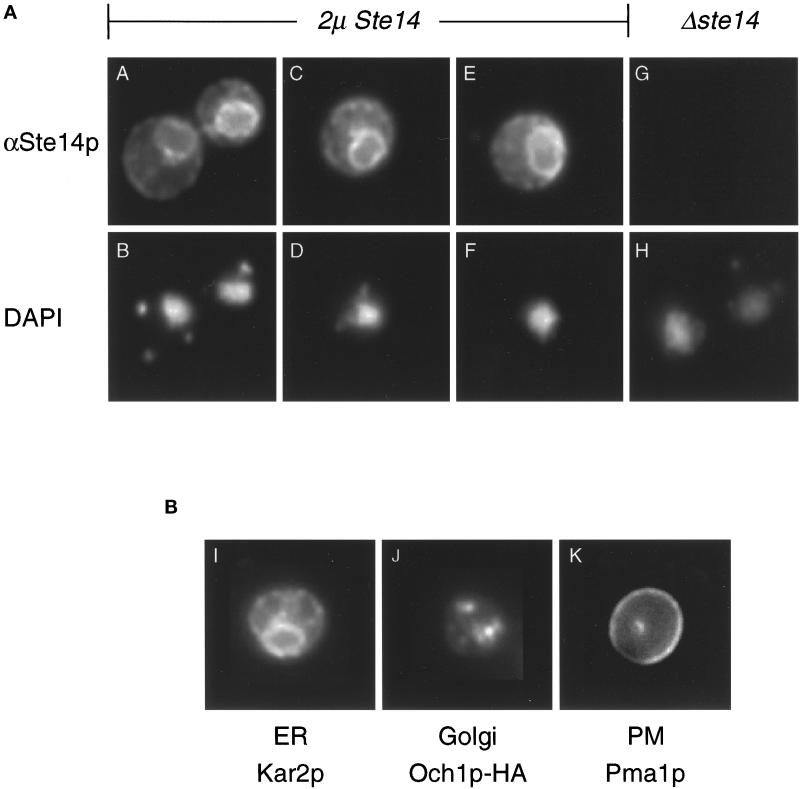
Figure 3.
Ste14p is localized to the ER membrane by immunofluorescence. (A) Immunofluorescence pattern of Ste14p. Indirect immunofluorescence of Ste14p was performed in Δste14-3 strains bearing either pSM1317 (2μ URA3 STE14) (panels A-F) or pRS316 (CEN URA3) (panels G and H). Cells were fixed with formaldehyde, spheroplasted, and probed with a 1:2000 dilution of anti-Ste14p antiserum (αSte14p) that had been depleted previously of nonspecific antibodies, followed by secondary decoration with Cy3-conjugated goat anti-rabbit antiserum (panels A, C, E, and G) and staining with DAPI (panels B, D, F, and H). The images in panels A, C, E, and G were exposed for equivalent amounts of time. (B) Immunofluorescence patterns for a group of marker proteins. The typical immunofluorescence staining pattern of an ER, Golgi, and plasma membrane marker is shown. Fixed and permeabilized cells from an SM1058 background expressing CEN URA3 plasmids or CEN URA3 OCH1::HA (SM3495) were stained with a 1:1000 dilution of anti-Kar2p antiserum (panel I), a 1:1000 dilution of anti-HA antiserum (panel J), or a 1:200 dilution of anti-Pma1p antiserum (panel K). The localization of Kar2p (ER), Och1p-HA (Golgi), and Pma1p (PM) shown here is consistent with previously observed localization data (Rose et al., 1989; Harris et al., 1994; Harris and Waters, 1996).

