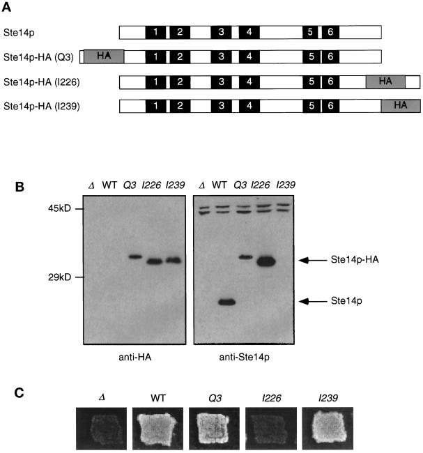
Figure 5.
Introduction of epitope tags into Ste14p. (A) Diagram showing the placement of the triple HA epitope tag in Ste14p. Putative membrane spans (solid bars) and the triple HA epitope (gray bars) are indicated and shown in approximate proportions. The epitope tag (HA) was placed at the N terminus (Q3), internally (I226), or at the C terminus of Ste14p (I239). (B) Detection of tagged and untagged Ste14p and Ste14p-HA by anti-HA and anti-Ste14p antisera. Crude cell extracts were subjected to 12.5% SDS-PAGE and transferred to nitrocellulose, and immunoblots were probed with either anti-HA antiserum or anti-Ste14p antiserum depleted previously of nonspecific antibodies as indicated. Lanes contain SM2926 (Δ), SM3185 (WT), SM3187 (Q3), SM3431 (I226), and SM3189 (I239). (C) Patch mating assay. Patches of the indicated MATa strains were replica plated onto a lawn of the MATα mating tester (SM1068) spread on an SD plate and incubated at 30°C for 2 d. Mating is detected by the growth of prototrophic diploids as indicated for WT, Q3, and I239. No mating is observed for Δste14 or I226.

