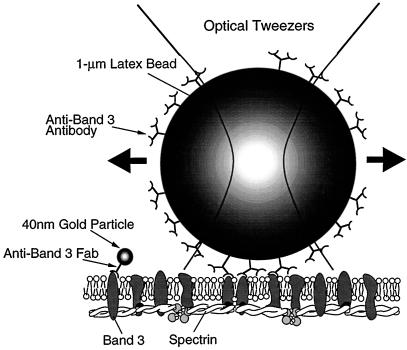
Figure 4.
Deformation of the membrane skeleton using optical tweezers. A 1-μm latex bead, coated with anti-band 3 IgG, bound multiple band 3 molecules, of which ∼30% are attached to the membrane skeleton. By dragging the latex bead, it was possible to deform the membrane skeleton, which can be observed by single particle tracking using gold particles specifically attached to spectrin on the cytoplasmic surface of the erythrocyte ghost membrane.

