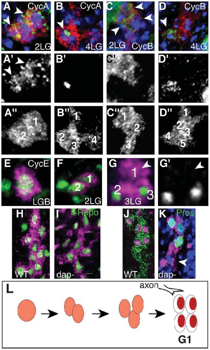
Fig. 6. Late segregation of Pros does not require asymmetric cell division.

LG are visualized with anti-βgal (red or magenta) in LG-lacZ reporter embryos. (A,B) Anti-CycA (green) is (A) cytoplasmic at 2-LG (arrowheads) and (B) nuclear at the 4-LG stage (arrowheads). (C,D) Anti-CycB is first cytoplasmic (arrowhead, C) and nuclear at the 4-LG stage (arrowhead, D). (E-G) Anti-CycE. CycE is in the LGB and 2-LG stage and it is first downregulated in one LG at the 3-LG stage (G is a 3-D rendered image). (H,I) Glial nuclei are visualized with anti-Repo (green) at stage 15. (H) Wild-type embryos. (I) dacapo mutant embryos (lines indicate LG cluster from one hemisegment) with four LG instead of the normal ∼10 at this stage (stage 15). (J,K) LG stained with Pros (green) at stage 16. (J) wild-type. (K) Despite having four LG instead of ten, Pros is downregulated in one posterior LG in dacapo mutants (white arrowhead) and present in anterior LG. DNA is stained with TOTO-3 in blue. (L) Diagram illustrating the onset of the first G1 in the LG lineage. Anterior is up. (A’-D’,G’) are single channel images of the cyclins. All images represent one hemisegment and the progeny of one glioblast.
