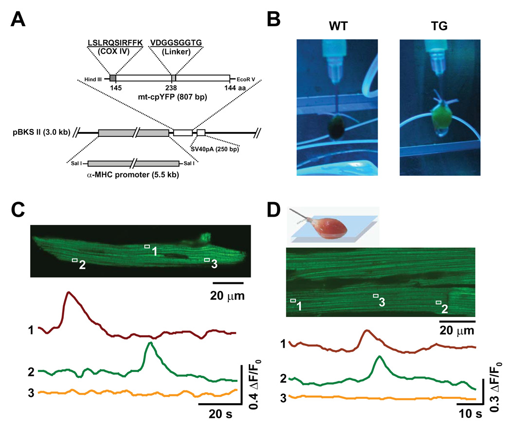
Figure 3. Superoxide Flashes in mt-cpYFP Transgenic Mice.
(A) Schematic of the cardiac-specific mt-cpYFP expressing vector used to generate mt-cpYFP transgenic mice. (B) Images of Langendorff-perfused hearts of wild type (WT) and mt-cpYFP transgenic mice (TG) under UV illumination. Note the green fluorescence of the TG heart. (C) Visualization of superoxide flashes in a freshly isolated ventricular myocyte from a TG mouse. Upper panel: Image of a representative myocyte. Lower panel: Time course of the mt-cpYFP signals (488 nm excitation) from mitochondria indicated in the image, showing two active and one quiescent mitochondria. Similar results were obtained in 12 myocytes from 3 TG mice. (D) Imaging of superoxide flashes in the Langendorff-perfused heart from a TG mouse. Upper panel: Illustration of experimental setting. Middle panel: 2-D image of cardiac myocytes in the myocardium of the beating heart from a TG mouse. Lower panel: time course of mt-cpYFP signals from two active and one quiescent mitochondria (box 3, illustrating absence of motion artifact). Similar results were obtained in 4 other hearts.

