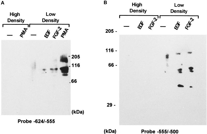
Figure 8.
Southwestern analysis of protein binding to PMA/PKC-responsive (A) and growth factor-responsive (B) regions. Thirty-five micrograms of nuclear proteins isolated from confluent (high-density cultures) or subconfluent (low-density) astrocytes incubated in control serum-free medium (−) or treated with 0.5 nM 18-kDa FGF-2, 5 nM EGF, or PMA (100 nM) for 24 h were resolved on SDS-PAGE gels and electroblotted to nitrocellulose membranes. The membranes were probed with 32P-labeled (1 × 10⁶ cpm/ml) −624/−555-bp (A) or −555/−500-bp (B) FGF-2 promoter fragment. The approximate molecular masses of the detected proteins were estimated by comparing with prelabeled protein standards. No protein binding was detected with 32P-labeled −453/−274-bp or −274/+11-bp FGF-2 promoter fragments.

