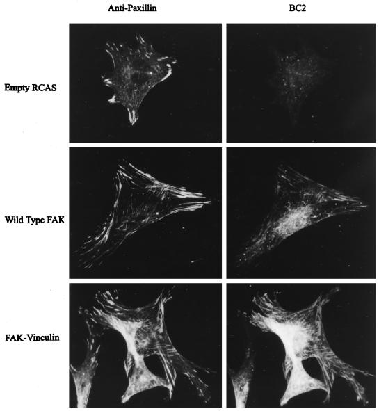
Figure 8.
Subcellular localization of the FAK/vinculin chimera. Cells transfected with empty RCAS (top) and cells expressing wild-type FAK (middle) or the FAK/vinculin chimera (bottom) were fixed and stained using purified BC2 (right). Cells were costained using a paxillin monoclonal antibody (left).

