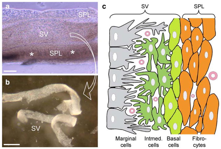
Fig. 1.
Structure of the cochlear lateral wall. a Isolated cochlear lateral wall (asterisks spiral prominence and the inferior region below the spiral prominence, SV stria vascularis, SPL spiral ligament). b The split stria vascularis. c Representation of the cochlear lateral wall (Intermed. intermediate). Bars100 μm (a), 0.5 mm (b)

