Fig. 2.
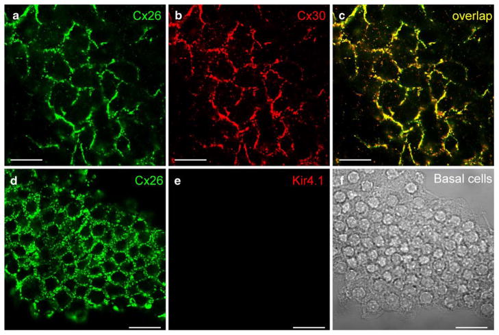
Cx26 and Cx30 labeling in the basal cell layer of the rat stria vascularis (SV). a, b Immunofluorescent images of double-labeling of the SV for Cx26 and Cx30. Confocal images show a honeycomb-like labeling pattern. c Merged image of a, b (yellow co-localization). d, e Double-immunofluorescent labeling of the basal cells for Cx26 and Kir4.1. Cx26 labeling shows a honeycomb-like pattern between the basal cells, but no labeling for Kir4.1 is visible. f Nomarski image of fields in d, e. Bars20 μm in (a–c), 40 μm (d–f)

