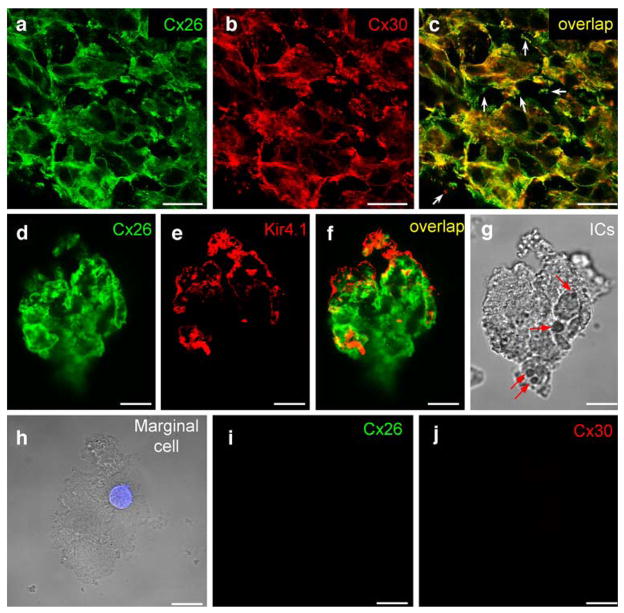
Fig. 3.
Immunofluorescent labeling for Cx26 and Cx30 in the stria intermediate cells and marginal cells. a–c Double immunofluorescent labeling for Cx26 and Cx30 in the intermediate cell layer of the SV of guinea pig (yellow co-localization, white arrows in c punctate labeling at the cell edge). d–g Immunofluorescent labeling for Cx26 and Kir4.1 in a dissociated intermediate cell group from rat. Both Cx26 and Kir4.1 show positive labeling (yellow co-localization, arrows in g black pigment granules visible in cytoplasm, ICs intermediate cells). h–j Negative immunofluorescent labeling for Cx26 and Cx30 in a marginal cell. The cell nucleus is revealed by DAPI staining (blue). No pigment granules are visible in the cytoplasm. Bars20 μm (a–c), 10 μm (d–j)

