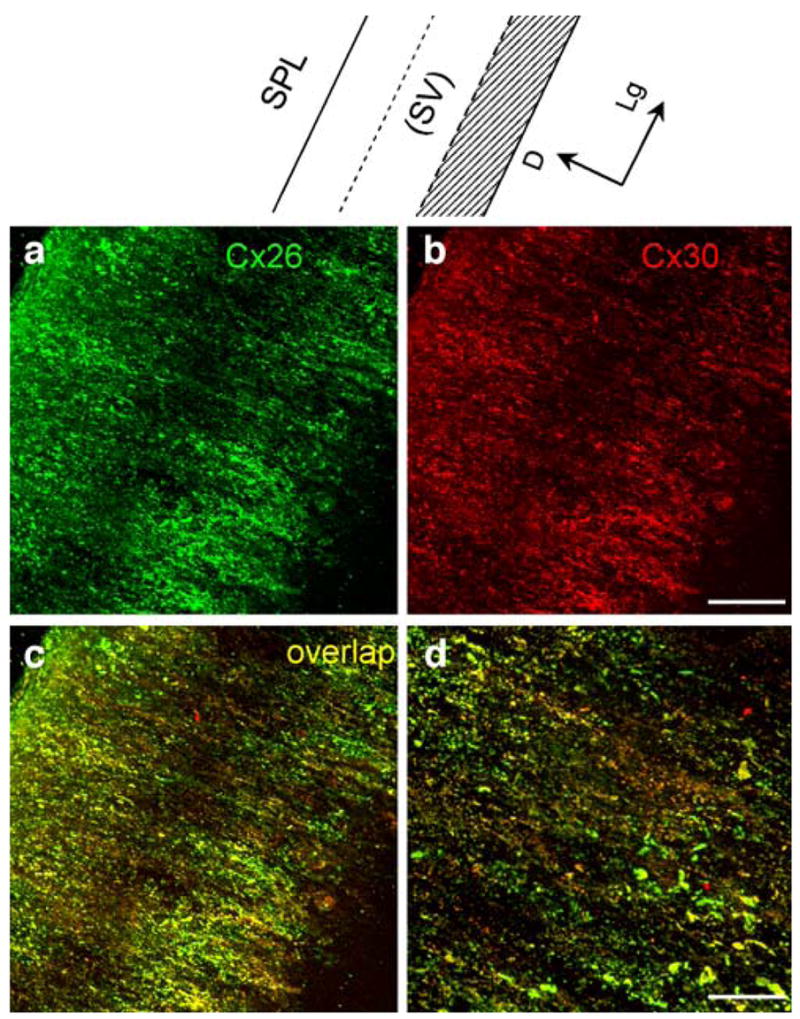
Fig. 4.

Double immunofluorescent labeling of the rat spiral ligament (SPL) for Cx26 and Cx30 (SV stria vascularis). a, b Confocal immunofluorescent images for Cx26 and Cx30 labeling. c Merged image of a, b (yellow co-localization). d Higher magnification image of c. Note that the SV had been removed from the SPL before staining. Top Representation of the SPL for orientation (shadowed area spiral prominence and the inferior region below the spiral prominence, D dorsal, Lg longitudinal). Bars 50 μm (a–c), 20 μm (d)
