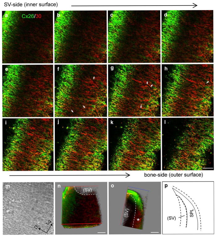
Fig. 5.
Cx26 and Cx30 labeling in serial confocal scanning sections of the separated rat spiral ligament (SPL) at the middle turn. a–l Sections were serially scanned from the inner surface (SV-side) of the SPL to its outer surface (bone-side) in 3.25-μm steps (green Cx26 labeling, red Cx30 labeling, yellow co-localization, white arrows in f–h “shadows” of blood vessels). m Nomarski image of d (D dorsal, Lg longitudinal). n, o Reconstructed three-dimensional images. Red, green, and blue axes represent the X, Y and Z axes, respectively. n View of the X-Y plane from a small angle in the Z axis. Note that the stria vascularis (SV) had been removed from the SPL before staining. o Orthogonal cross-sectional view at the X-Z plane (asterisk region beneath the SV with little Cx26 labeling). Bars50 μm. p Representation of the orthogonal cross-sectional view at the X-Z plane

