
Figure 1.
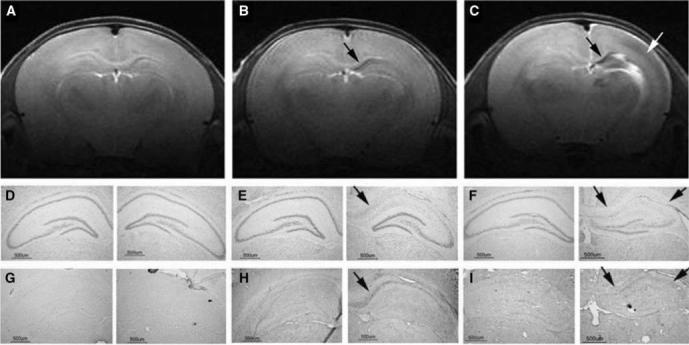
Post-hypoxic–ischemic (HI) hippocampal injury. Magnetic resonance imaging and brain sections from three iron-deficient rats obtained 1 week after a unilateral HI injury of 15, 30, or 45 mins on postnatal day 14 showed no hippocampal injury (A, D, and G), neuronal injury and demyelination limited to CA1 hippocampal subarea (arrows in B, E, and H) and injury involving the whole hippocampus (black arrows in C, F, and I) and the adjacent parietal cortex (white arrow in C) on the HI side, respectively. The distribution of rats with these injuries in the two dietary groups is given in Table 1. (15 μm coronal brain sections, Nissl histochemistry, and immunohistochemistry for degraded myelin basic protein on adjacent brain sections. See text for details of RARE image acquisition).