
Figure 6.
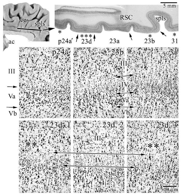
Case #6 was cut in the horizontal plane and reacted for NeuN and shows the features of transition from area p24a’ to area 31. The macrophotograph has an extension of retrosplenial cortex (RSC) from the callosal sulcus along the ventral border of the cingulate gyrus and marks the end of the splenium of the corpus callosum in more ventral sections. Each asterisk in this latter section shows where microphotographs were taken and the arrows indicate the borders between adjacent areas. In addition to the architectures of areas in this section, the bottom three microphotographs are of area 23d. The long double arrows delimit layer IV and adjacent layers and the double asterisks show points at which there is an intermingling of large layer IIIc and Va pyramidal neurons as is characteristic of dysgranular cortices. spls, splenial sulcus.