
Figure 10.
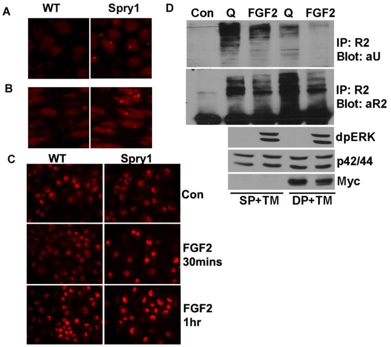
Overexpression of Spry1 resulted in accumulation of FGFRs in chondrocytes possibly through inhibiting degradation of FGFRs. Immunofluorescence microscopy showed an increase of FGFR3 immunoreactivity in the reserve zone (A) and proliferating zone (B) in Spry1;Col2-Cre growth plates. (C) Primary chondrocytes from Spry1;CAGGCre-ER™ embryos were induced to express Spry1 by the addition of 1 μM tamoxifen for 48 h, followed by the addition of FGF2 for the indicated times. Immunofluorescence staining for FGFR2 demonstrated a more rapid accumulation of FGFR2 in nucleus when Spry1 was overexpressed. (D) Primary chondrocytes from Spry1;CAGGCre-ER™ embryos (DP) or Cre-negative controls (SP) were treated with 1 μM tamoxifen (TM) for 48 h. Chondrocytes were then either left unstimulated (Q) or stimulated with FGF2 for 15 min. Immunoprecipitation with FGFR2 antibodies showed FGF2 stimulation induced degradation of FGFR2, and overexpression of Spry1 inhibited ubiquitination of FGFR2 in the quiescent state, and resulted in accumulation of FGFR2.