
Figure 2.
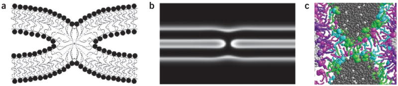
The stalk is the key intermediate in most of the theoretical models developed with the continuous and the simulation approaches. (a) Stalk structure computed by analysis of bending, splay and tilt of the lipid molecules in the membrane monolayers with the elastic model (continuous approach)27. (b) Stalk structure computed by the self-consistent field model (continuous approach)48. Light regions indicate the areas of head groups of the bilayer. (c) Stalk structure ‘observed’ by molecular dynamics simulation of the fusion between liposomes composed of dipalmitoyl phosphatidylcholine and palmitic acid using an atomistically detailed model. Water molecules (gray) and head group atoms of the lipids are depicted as spheres; tails are shown as bonds, with gray used to distinguish water molecules originating on different sides of the fusing membranes. The coloring also distinguishes between lipid molecules coming from different leaflets of the bilayers: dipalmitoyl phosphatidylcholine molecules in the inner or outer leaflets (green and purple), and palmitic acid in the inner or outer leaflets (cyan or magenta, respectively).