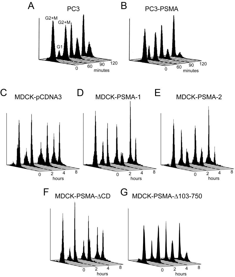
Figure 3.
Analysis of the cell cycle progression in PSMA expressing cells after release of the mitotic block. Cells were blocked in mitosis as described in Materials and Methods. The mitotic block was released for 60, 90, and 120 minutes for PC3 clones (A, B) and 2, 4, and 8 hours for MDCK clones (C-G) and analyzed for cell cycle progression. Profiles shown are representative of the data shown in Table 1.

