
Figure 6.
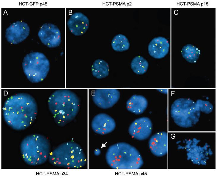
PSMA expressing human cells are genetically unstable. HCT-PSMA and HCT-GFP cells were analyzed by multicolor FISH with Chromosome 3 labeled with Spectrum Red (CEP3), Chromosome 7 with Spectrum Green (CEP7), Chromosome 17 with Spectrum Aqua (CEP 17), and 9p21 region (p16 gene) with Spectrum Gold. Control HCT-GFP cells at passage 45 (A), and HCT-PSMA cells at passage 2 (B), are diploid whereas HCT-PSMA cells at passage 15 (C), 34 (D), and 45 (E) show aneuploidy. Micronucleus formation (E, arrow and F) and abnormal metaphase, (G) in HCT-PSMA cells at passage 45 are shown.