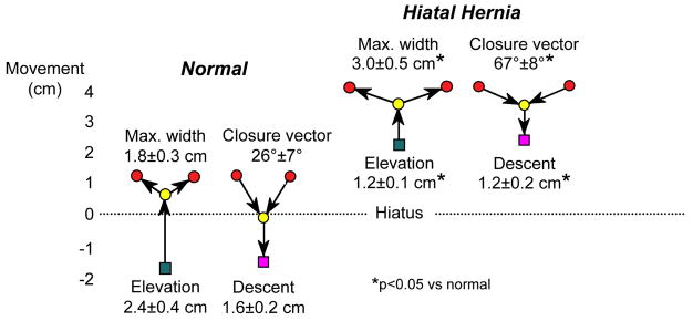
Figure 3.
Axial mobility of the EGJ during swallow in normal controls and patients with type I hiatus hernia demonstrated by fluoroscopically tracking the movement of endoscopically place endoclips at the squamocolumnar junction. Clip movements are referenced to a point on the vertebral column. The green square represents the position of the SCJ prior to swallow, almost 2 cm distal to the center of the hiatus in the normal controls and 2 cm above it in the case of the hernia patients. With the initiation of swallow, the clip elevates to the point indicated by the yellow circle before luminal opening occurs. The red circles then indicate the maximal diameter of opening achieved, significantly wider in the case of the hernia patients, consistent with the reduced compliance demonstrable in these patients. During closure the angle of descent is steeper with the normals than with the hernia patients and the final position (magenta square) is essentially the same as prior to the swallow. Laxity and loss of elasticity of the phrenoesophageal membrane is suggested by two observations: 1) the attenuated axial movement of the SCJ in the hernia patients and 2) the flattening of the closure vector in the hernia patients suggesting diminished elastic recoil as the longitudinal muscle of the esophagus relaxes post-swallow. Modified from Kahrilas, PJ, Wu, S, Lin, S, et al. Attenuation of esophageal shortening during peristalsis with hiatus hernia. Gastroenterology 1995;109:1818.

