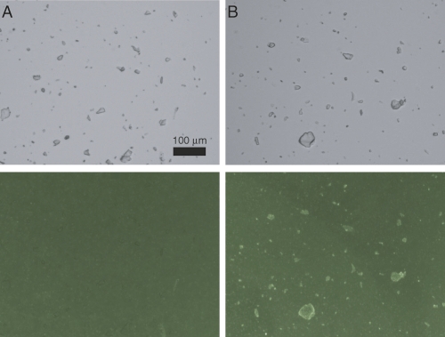
Figure 4.
Fluorescent lipid binding to insulin crystals. Microcrystals of human insulin were incubated in CZ6 buffer with 0.13 mM DOPG liposomes containing 0.25% NBD-DOPE in the absence (A) or presence (B) of 7 μM rat IAPP. Crystals are clearly visible in phase contrast mode (top). Lipid binding is evidenced by bright crystal surfaces when viewed with a FITC fluorescence filter (bottom). Scale bar (100 μm) applies to all panels.

