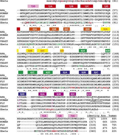
Figure 3.
Sequence alignment of RACK1A from A. thaliana (GI:30685669) versus RACK1 from Homo sapiens (GI:83641897), Drosophila melanogaster (GI:24582618), and S. cerevisiae (GI:6323763), as well as a structural alignment with respect to the crystal structure of Gβ in complex with Gα and Gγ (PDB ID code 1GP2). Secondary structural elements in RACK1A are shown above the alignment and colored according to the ribbon diagram in Figure 1B. Identity between RACK1A from A. thaliana and human RACK1 versus the other proteins is listed at the end of the alignment. Residues conserved in all RACK proteins shown are marked with an asterisk below the residue. Residues in Gβ that are structurally similar to residues in RACK1A are capitalized. Residues of Gβ that line the interface with Gγ interface are colored red. Residues lining the interface with Gα are colored green. Tyrosine RACK1 residues predicted to be phosphorylated are colored purple and the predicted sumoylation site of human RACK1 is underlined.

