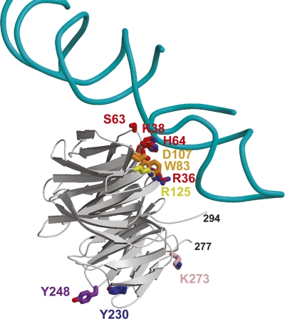
Figure 6.
Ribbon diagram of the model of RACK1 binding to the 80S ribosome complex, based on the cryo-EM structure (Sengupta et al. 2004). Shown are the H39 and H40 of rRNA from the 40S subunit and RACK1A. To make this figure, RACK1A was superimposed on the model of RACK1 created from Gβ (PDB ID code 1TRJ). Residues colored red, orange, and yellow are conserved residues from conserved site 1 of RACK1A. The purple tyrosine residues are the sites of putative Src phosphorylation at the second conserved site. The pink lysine is residue 273, the proposed sumoylation site. This figure suggests conserved region 1 could interact with the ribosomal RNA and conserved region 2 could recruit Src to the ribosome.

