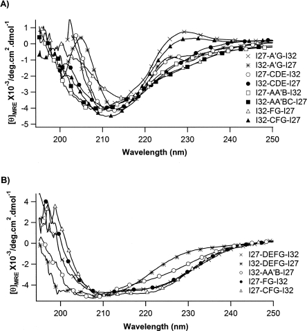
Figure 2.
Far-UV CD spectra for the recombined hybrid Ig domains. (A) CD spectra of eight engineered hybrid Ig proteins that show predominantly β-sheet structure. These spectra are characterized by single minima between 210 and 218 nm. (B) CD spectra of engineered hybrid Ig domains that show secondary structures deviating from typical β-sheet structure. The CD spectra of these proteins show a broad negative peak between 208 nm and 222 nm, with two minima at ∼208 nm and ∼222 nm, suggesting the formation of an α-helical structure.

