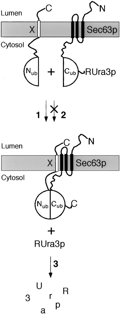
Figure 1.
The split-Ubiquitin technique and its application to the analysis of membrane proteins using a metabolic marker. Cub-RUra3p was linked to the C terminus of Sec63p, and Nub was linked to the N terminus of the membrane protein X. Pathway 1: Nub is coupled to a protein that binds to Sec63p. The complex brings Nub and Cub into close proximity. Nub and Cub reconstitute the quasi-native Ub that is cleaved by the Ub-specific proteases to release RUra3p from Cub. The cleaved RUra3p is targeted for rapid destruction by the enzymes of the N-end rule (3) to yield cells that are uracil auxotrophs and 5-FOA resistant. Pathway 2: Nub is linked to a protein that does not bind to Sec63p. The two fusion proteins do not improve the reconstitution of Nub and Cub into the quasi-native Ub. Thus, RUra3p stays linked to Sec63-Cub, and the cells are uracil prototrophs and 5-FOA sensitive.

