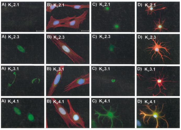
Fig. 5.
Immunoreactivity for Kir 2.1, 2.3, 3.1, and 4.1 in STTG1 cells and mature spinal cord astrocytes. A: STTG1 cells stained with the four different Kir antibodies. B: Merged image of STTG1 cells with the four Kir antibodies (green), phalloidin (red), and DAPI (blue). C: Spinal cord astrocytes at 9 days in culture (DIC) with the four Kir antibodies. D: Merged image of the spinal cord astrocytes with the four Kir antibodies (green), with GFAP (red) and DAPI (blue). Scale bar = 20 μm.

