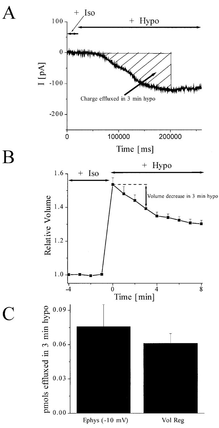
Fig. 5.
Magnitude of chloride effluxed through hypotonically activated chloride channels accounts for mols of solute effluxed during volume regulation of astrocytes. A: Representative example of gap-free currents obtained at a -10 mV holding potential during 3-min hypotonic challenge. Note that the -10 mV holding potential corresponds to an 8.7-mV hyperpolarization from the calculated ECl. The total charge effluxed during this time was calculated by integrating the area under the curve (hatched). B: Normal volume regulation plotted for 10 min following exposure to hypotonic challenge. The volume decrease within the 3 minutes of interest is shown with an arrow. C: Comparison of pmols effluxed during 3-min hypotonic challenge in gap-free electrophysiological recordings at -10 mV holding potential and during volume regulation. An explanation of the calculations is given in the Results section.

