
Fig. 7.
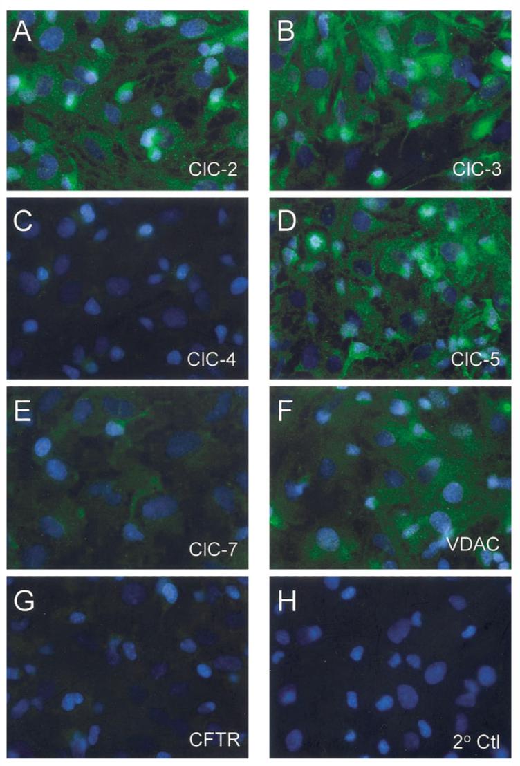
Immunocytochemical detection of previously cloned chloride channels in astrocytes. Cultured rat cortical astrocytes were evaluated for the presence of chloride channels proteins that correlated with those detected at the RNA level in Fig. 6. These included ClC channels (A-E), VDAC (F), and CFTR (G). The strongest staining was visible for ClC-2 (A) and ClC-3 (B). Strong staining was also visible for ClC-5 (C) and VDAC (F). Weak staining was visible for ClC-7 (E) and very weak staining was visible for ClC-4 (C) and CFTR (G). Note that cells were not permeabilized and that staining represented protein localized to the cell membrane. Control stainings were performed by exclusion of primary antibody (H).