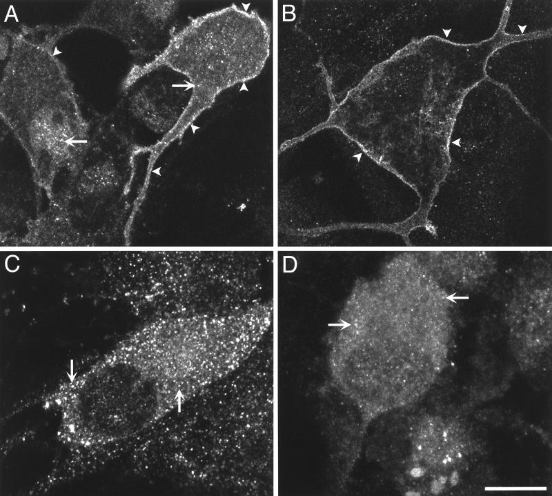
Figure 7.
Confocal images showing the localization of immunoreactive NK1-R, Gαq/11, GRK-2, and β-arrestin-1 and -2 in myenteric neurons not exposed to SP. (A) Localization of the NK1-R at the plasma membrane of the soma and neurites (arrowheads) and in some vesicles (arrows). (B) Localization of Gaq/11 at the plasma membrane of the soma and neurites (arrowheads). (C) Punctate and cytosolic localization of GRK-2 (arrows). (D) Punctate and cytosolic localization of β-arrestin-1 and -2 (arrows). Bar, 7.5 μm (A and B), 10 μm (C and D).

