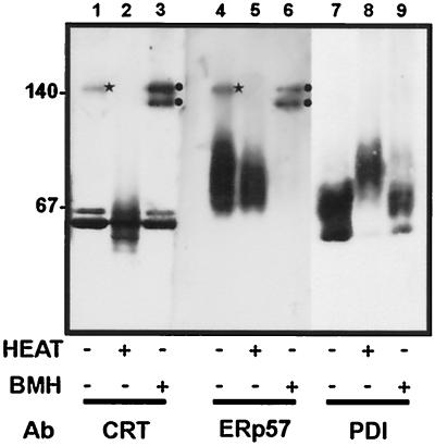
Figure 2.
Native gel analysis of ER protein complexes. The lumenal contents of canine pancreatic microsomes were separated by blue native gel electrophoresis and then transferred to PVDF. The proteins were detected by immunoblotting with antisera raised against calreticulin (CRT, lanes 1–3), ERp57 (lanes 4–6), and PDI (lanes 7–9). The samples were untreated (lanes 1, 4, and 7); heated to 95°C before electrophoresis (lanes 2, 5 and 8), or treated with 1 mM BMH before isolation of the lumenal contents (lanes 3, 6, and 9).

