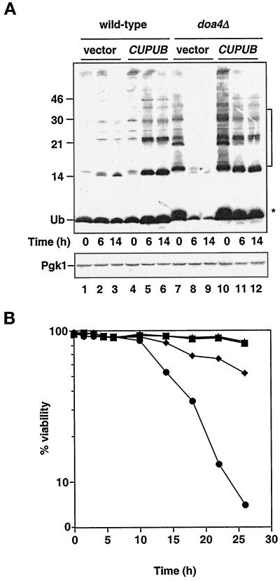
Figure 1.
Characteristics of stationary phase doa4Δ cells. (A) Analysis of ubiquitin levels in wild-type and doa4Δ cells at distinct stages in the growth cycle. Lanes 1–3, wild type (vector); lanes 4–6, wild type (YEp96); lanes 7–9, doa4Δ (vector); lanes 10–12, doa4Δ (YEp96). At the indicated time points, extracts for immunoblot analysis were made from the same yeast cultures that were used in B. Anti-ubiquitin antibody binding was detected by enhanced chemiluminescence, whereas anti-Pgk1 antibody binding was followed by [125I]protein A. The * and bracket indicate the positions of ubiquitinated species that accumulate in doa4Δ cells. The slight increase in ubiquitin (Ub) levels in wild-type cells carrying YEp96 at 6 h is most likely caused by upregulation of the CUP1 promoter in response to nutrient starvation (Tamai et al., 1994). The position of size standards (in kilodaltons) is indicated on the left. (B) Viability of wild-type and doa4Δ cells in the presence and absence of exogenously supplied ubiquitin. Rates of survival of wild-type (MHY501) and congenic doa4Δ (MHY623) cells carrying pES12 (vector) or YEp96 (CUP1UB) were measured by propidium iodide staining. Viability measurements were begun when yeast cultures were in logarithmic growth at an OD600 of ∼0.5, ∼10 h before growth levels off. The symbols denote the following strains: wild type (vector; ▪), wild type (CUP1UB; ▴), doa4Δ (vector; ●), and doa4Δ (CUP1UB; ♦).

