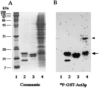
Figure 2.
Far Western blot analysis with 32P-labeled GST-Act3p(Ins II). Marker proteins (lane 1), chicken histone fraction enriched in H3 and H4 (lane 2), purified chicken histones H2A and H2B (lane 3), and the chromatin fraction prepared from isolated yeast nuclei (lane 4) were electrophoresed on SDS-polyacrylamide gels. The gels were subjected to staining with Coomassie brilliant blue R-250 (A) and to far Western blot analysis with 32P-labeled GST-Act3p(Ins II) (B). Radioactivity of the 32P-labeled peptide bound to proteins on the membrane were detected with BAS2000 bio-image analyzer. An arrow and an arrowhead to the right of B mark the positions of a 17-kDa band and a 30-kDa band in lane 4, respectively.

