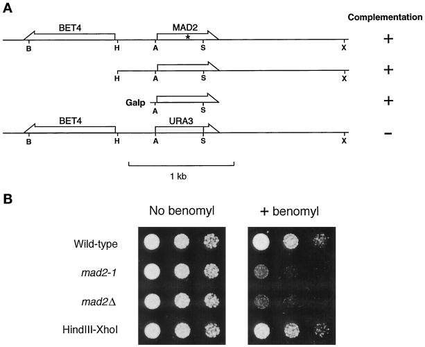
Figure 1.
Identification of MAD2. (A) Relative position of MAD2 and BET4 and the ability of various constructs to rescue mad2-1. The mutation in mad2-1 is marked with an asterisk. The positions of the following restriction enzyme recognition sites are indicated: B, BamHI; H, HindIII; A, ApaI; S, ScaI; X, XhoI. (B) Benomyl sensitivity of mad2 mutants. Cells were spotted onto either a YPD plate (left panel) or a YPD plate containing 7.5 μg/ml benomyl (right panel). Cells were diluted 10-fold from the corresponding spot on the left. Yeast strains are indicated on the left. The HindIII–XhoI fragment upstream of BET4 fully rescued the benomyl sensitivity of mad2-1 when carried on a CEN plasmid.

