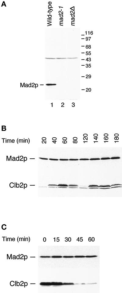
Figure 2.
(A) Specificity of anti-Mad2p antibody. The antibody recognizes a 24-kDa protein in wild-type cells (lane 1) that is not detectable in mad2-1 (lane 2) and mad2Δ (lane 3) strains. The migration of molecular size standards is indicated on the right. (B) Mad2p level and its mobility on SDS-PAGE stay constant throughout the cell cycle. Cells arrested at G1 with α-factor were released from the arrest for the time indicated. Cell lysates were immunoblotted with an anti-Mad2p antibody (upper panel) or with an anti-Clb2p antibody. (C) Mad2p levels and gel mobility remain unchanged at the metaphase to anaphase transition. Cells arrested at mitosis with benomyl and nocodazole were released from the arrest for the time indicated on top. Cell lysates were immunoblotted with an anti-Mad2p antibody (upper panel) or with an anti-Clb2p antibody (lower panel).

